
Shoulder Instability: Causes, Types, and Treatment Options
The shoulder is the most mobile—and least stable—joint in the human body, allowing movement in multiple planes. Shoulder instability occurs when the head of the humerus is unable to remain properly centered within the glenoid fossa during movement.
Instability exists on a spectrum. In some cases, the humeral head partially translates beyond normal limits while still maintaining contact with the glenoid—this is known as a subluxation. In more severe cases, the humeral head moves completely beyond the rim of the glenoid, resulting in a dislocation. When the surrounding anatomical structures can no longer control this motion, symptoms such as pain, weakness, and loss of function often develop.
The glenohumeral joint relies on a complex balance of static and dynamic stabilizers. When one or more of these structures are compromised—through trauma, repetitive stress, or connective tissue laxity—shoulder instability can develop.
Common injuries associated with shoulder instability include:
Subluxation
Dislocation (with or without reduction)
Rotator cuff tears
Hill-Sachs lesions
Bankart lesions
Anatomy of Shoulder Stability
Static Stabilizers
These structures provide passive stability to the shoulder:
Glenoid labrum
Joint capsule
Glenohumeral ligaments
Negative intra-articular pressure
Dynamic Stabilizers
These muscles actively control shoulder motion:
Rotator cuff: supraspinatus, infraspinatus, teres minor, subscapularis
Periscapular muscles: serratus anterior, rhomboids, trapezius, latissimus dorsi, pectoralis minor
Deltoid
Proper coordination between these stabilizers is essential for maintaining shoulder health.
Types of Shoulder Instability
Anterior Shoulder Instability
Anterior instability is the most common type, accounting for approximately 95% of instability events. It occurs when the humeral head translates forward (anteriorly), often during positions of abduction and external rotation.
This can result from:
Traumatic events (falls, collisions)
Repetitive overhead movements
Anterior instability is more common in younger individuals due to ligamentous laxity, while adults over age 40 often experience instability related to rotator cuff tears.
Sports with higher risk include:
Baseball
Volleyball
Swimming
Posterior Shoulder Instability
Posterior instability is less common, accounting for approximately 2–5% of cases, but is still seen in active populations. It typically occurs when the shoulder is placed in flexion, adduction, and internal rotation.
It may result from:
Repetitive microtrauma
High-force macrotrauma
Posterior instability is more frequently seen in males ages 20–30, particularly in:
Weightlifting
Contact sports such as football
Multidirectional Shoulder Instability
Multidirectional instability involves instability in two or more planes (anterior, posterior, and/or inferior). It is most common in individuals aged 20–30 years and is often associated with repetitive microtrauma from overhead activities.
It may also be linked to generalized joint laxity seen in conditions such as:
Ehlers-Danlos syndrome
Marfan syndrome
Osteogenesis imperfecta
Hypermobility spectrum disorders
Common Clinical Signs and Symptoms
Shoulder pain (anterior, posterior, or deep ache)
Weakness or fatigue
Popping, clicking, or catching sensations
Neurological symptoms (tingling or burning)
Crepitus
Excessive range of motion
Poor dynamic control
Treatment Options for Shoulder Instability
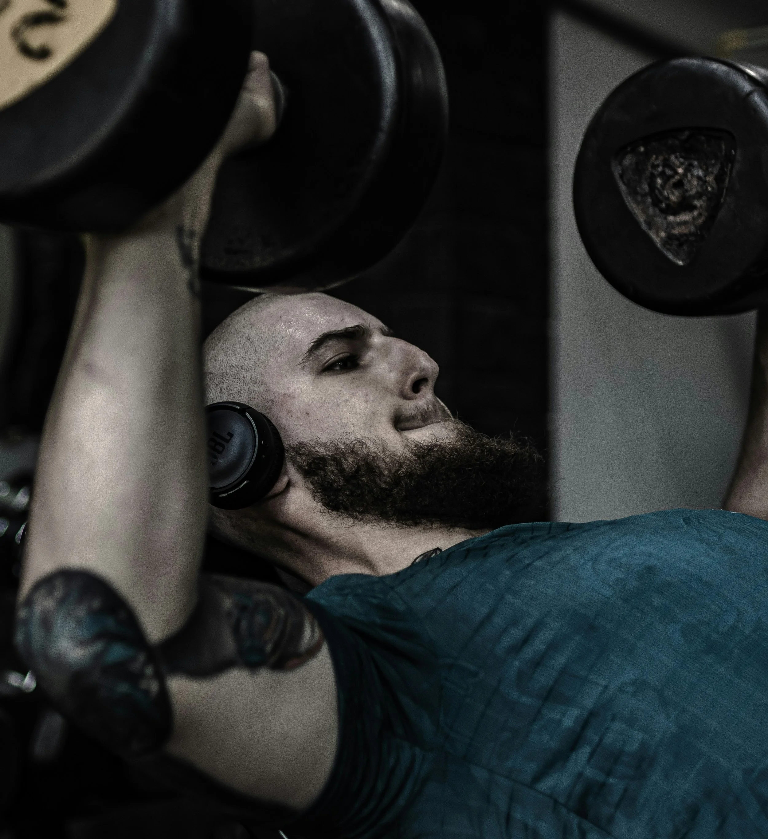
Physical therapy is typically the first line of treatment for shoulder instability and has been shown to be highly effective, particularly for:
Adults over age 40
Individuals not involved in high-demand overhead sports
Conservative management focuses on:
Scapular posture and control
Strengthening dynamic stabilizers
Proprioception and neuromuscular control
Functional and sport-specific training
Research supports that a comprehensive, individualized rehab program can significantly reduce symptoms and often help patients avoid surgery.
For younger athletes involved in high-demand overhead or contact sports, surgical intervention may be recommended after an acute dislocation due to a higher risk of recurrent instability. Following immobilization, physical therapy is essential to restore function and safely return to sport.
Examples of Beneficial Exercises
Plank and single-arm plank
Side plank variations
Plank drags
High plank on unstable surfaces
Isometric internal and external rotation walkouts
Prone T’s, W’s, and Y’s
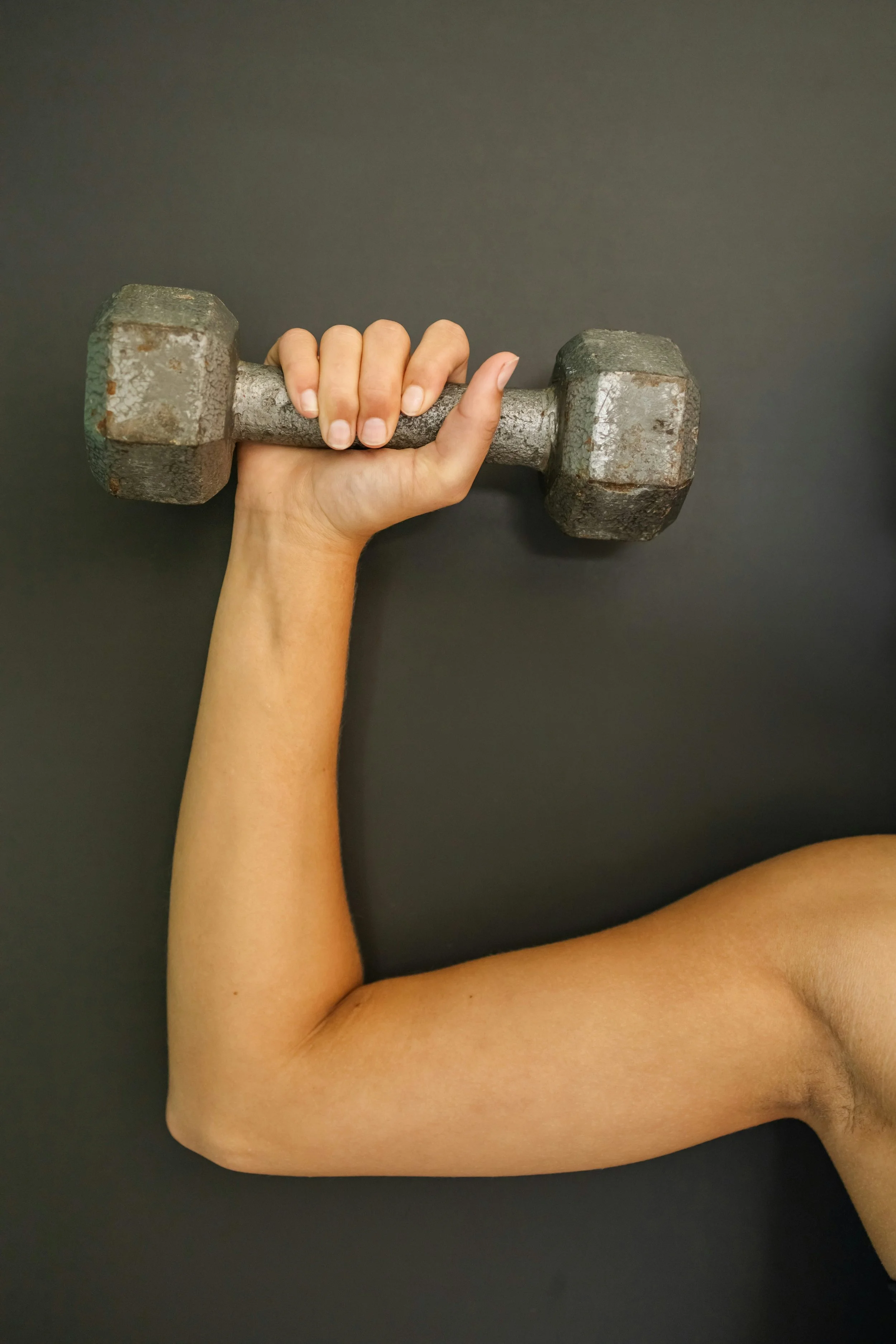
Carries (farmer’s, waiter’s, overhead)
PNF diagonal patterns
Jobe’s rotator cuff exercises
Exercise selection should always be tailored to the individual’s symptoms, goals, and activity demands.